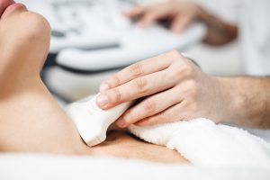
Vescular Ultrasound
24 weeks (adjustable for full-time or part-time schedules).
Format: Lectures, hands-on labs, case reviews, and assessments
Module 1: Introduction to Vascular Ultrasound
Overview of vascular ultrasound and its clinical applications.
Equipment and instrumentation: transducers, Doppler settings, image optimization.
Sonographer responsibilities, patient safety, and ergonomics.
Module 2: Vascular Anatomy & Physiology
Arterial and venous anatomy of the cerebrovascular, peripheral, and abdominal systems.
Hemodynamic: pressure, resistance, flow characteristics.
Normal vs abnormal flow patterns.
Module 3: Cerebrovascular Ultrasound
Anatomy of carotid and vertebral arteries.
Protocols for scanning the carotid arteries.
Plaque characterization and stenosis grading.
Spectral Doppler analysis and velocity criteria.
Case studies: TIA, stroke, and asymptomatic carotid disease.
Module 4: Peripheral Arterial Ultrasound (Upper & Lower Extremities)
Arterial anatomy of the limbs.
Indications for arterial exams: claudication, rest pain, ulcers.
Protocols: duplex imaging, waveform analysis, segmental pressures.
Stenosis and occlusion detection.
Post-surgical follow-up (bypass grafts, stents) .
Module 5: Peripheral Venous Ultrasound (Upper & Lower Extremities)
Venous anatomy: superficial, deep, and perforator systems.
Techniques for venous reflux and DVT evaluation.
Compression manoeuvres and augmentation.
Chronic venous insufficiency: CEAP classification.
Case studies: acute vs chronic DVT, varicose veins.
Module 6: Abdominal Vascular Ultrasound
Aorta and iliac arteries: aneurysms, dissections, stenosis.
Renal artery Doppler: indications, protocols, resistive index.
Mesenteric circulation: ischemia evaluation.
Inferior vena cava and hepatic vasculature.
Liver Doppler and portal hypertension assessment.
Module 7: Doppler Principles and Image Optimization
Colour, Power, and Spectral Doppler.
Aliasing, angle correction, and velocity measurements.
Optimizing settings for accurate diagnosis
Troubleshooting common Doppler artifacts.
Module 8: Case Interpretation and Clinical Correlation
Integrating clinical data with ultrasound findings.
Writing effective preliminary reports.
Reviewing real cases across all vascular regions.
Hands-on scanning labs and image review sessions.
Module 9: Professional Practice and Quality Assurance
Documentation standards and reporting guidelines.
Infection control and patient care.
Accreditation and credentialing (e.g., SVT, RVT, RVS) .
Continuing education and career development.
Assessment & Evaluation
Weekly quizzes and assignments.
Midterm and final written exams.
Practical skills assessment and scanning competency check-offs
Final case-based presentation or portfolio
Course Completion Outcomes
Upon completing the course, students will be prepared to:
Perform high-quality vascular ultrasound exams.
Interpret and document findings accurately.
Support clinical decision-making through detailed vascular assessments.
Sit for professional credentialing exams in vascular ultrasound.
Perform true-to-life patient examinations
Scan in real-time from the clavicle to the pelvis with seamless transition between cardiac and abdominal probes...
Key Features of the Course
Hands-on Scanning Sessions: Live practice on standardized patients/models. Case-Based Learning: Real-world pathologies with expert interpretation. Interactive Q&A: Troubleshooting common pitfalls in image acquisition. Certification: Accredited competency assessment
Six Months Vescular Ultrasound Course
Enroll in our One-Year Abdominal Ultrasound Course to enhance your expertise in diagnostic imaging. Visit our course program to explore how this valuable training can advance your medical career.