Abdominal Ultrasound
Course Overview
This intensive program equips sonographers, radiologists, and clinicians with advanced skills in abdominal and small parts ultrasound. Participants will master anatomical recognition, hemodynamic principles, and scanning protocols through theory lectures, case studies, and live demonstrations.
Detailed Course Contents
1. Foundations of Abdominal Ultrasound
General Information & Hemodynamic
Physics of ultrasound (knobology, artifacts, Doppler principles)
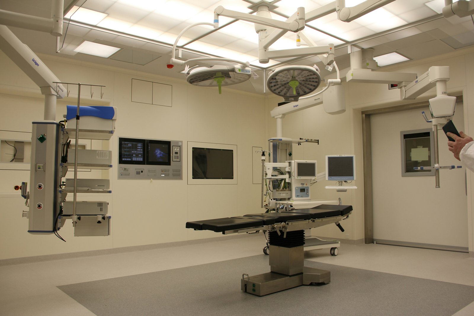
Patient positioning and ergonomics
Normal vs. pathological hemodynamic
2. Core Abdominal Organ Imaging
A. Abdominal Vasculature
Aorta, IVC, portal vein, and mesenteric vessels
Techniques for detecting aneurysms, stenosis, and thrombus
B. Liver Ultrasound: Segmental anatomy, lobes, and vasculature
Protocols for cirrhosis, fatty liver, and metastases
C. Gallbladder & Biliary Tree: Sonographic Murphy’s sign, sludge, stones, and choledochal cysts
Postprandial scanning techniques
D. Spleen: Size measurement, trauma assessment, and infarcts
E. Pancreas: Head/body/tail differentiation, pancreatitis, and pseudocysts
F. Urinary System: Kidneys (cortical thickness, hydronephrosis), bladder, and ureteral jets
Renal artery stenosis evaluation
3. Small Parts & Specialised Imaging
Prostate & Testicular Ultrasound
Transrectal vs. transabdominal approaches
Torsion, varicoceles, and tumour detection
Thyroid & Neck Ultrasound
Nodule characterization (TI-RADS), parathyroid glands
Miscellaneous Structures
Appendix, hernia evaluation, and paediatric considerations
Target Audience: Radiologists, Sonographers, ER Physicians, Internists.
Why This Course?
Covers both routine and complex cases with protocol standardization.
Emphasizes diagnostic accuracy and reporting standards.
Aligns with ASE/ACR guidelines for clinical relevance.
Why Enrol in This Course?
Boost Clinical Competence – Gain expertise in ultrasound scanning with real-world case studies.
Hands-On Learning – Practice on live models and simulators with 1:1 expert feedback
Earn Accreditation – Receive a certificate of completion recognized by leading medical institutions.
Stay Ahead – Learn latest protocols (ASE/ACR guidelines) and emerging techniques.
Course Highlights
Interactive Lectures – Simplified breakdown of complex concepts (e.g., Doppler physics, artifact troubleshooting).
Live Demos & Scanning Labs – Rotate through stations covering 9+ key organs Pathology Recognition – Differentiate benign vs. malignant findings (thyroid nodules, renal masses).
AI & Ultrasound – Bonus session on AI-assisted diagnostics!
Testimonials
“This course transformed my scanning speed and accuracy—especially in tricky pancreatic cases!”
– Dr. Sarah K., Radiologist
“Finally, a program that makes hemodynamics easy to understand!”
– James L., Senior Sonographer
By learning Abdominal Ultrasound module, we be able to:
Develop a thorough understanding of the regional surface anatomy of the abdomen, which serves as the foundation for effective ultrasound scanning. You will learn to properly position patients before a scan, ensuring the best possible image quality, and master the art of optimizing ultrasound control settings to enhance imaging. The module also covers recognizing common pathologies associated with key abdominal organs, including the liver, biliary tree, spleen, abdominal aorta, pancreas, kidneys, and bladder. By the end of this course, you will confidently identify normal anatomy, anatomical variants, and distinguish them from pathological findings. This includes detailed insights into the gallbladder, biliary tree, and the ability to review management algorithms for focal splenic lesions.
Additionally, the module delves into recognizing the different types of aortic aneurysms and identifying pancreatic anatomy and abnormalities using ultrasound. You will also gain a clear understanding of optimal techniques for liver examination, identifying both normal and abnormal findings, as well as comprehending the kidneys' anatomical position and the distinctions between normal variants and pathological appearances. The course emphasizes the importance of using the appropriate equipment to achieve high-quality imaging of the urinary bladder, understanding its normal anatomy, and identifying common pathologies.
Beyond these core areas, you will explore the ultrasound appearances of bowel and lymph nodes and learn to recognize conditions such as appendicitis, intussusception, ascites, and lymphadenopathy. A significant part of the training focuses on the FAST (Focused Assessment with Sonography in Trauma) scan, including the organs visible in its four key views, identifying free fluid, and understanding the limitations and potential pitfalls of the procedure. The module also covers the anatomy and postoperative complications associated with orthotopic transplanted livers and renal transplants, equipping you with the knowledge to perform thorough transplant assessments. Furthermore, you will study the anatomy of the chest and pleural cavity, gaining the ability to identify sonographic features of pneumothorax, pulmonary edema, and pleural fluid.
Finally, this comprehensive module will guide you through the limitations of abdominal ultrasound imaging and provide you with the skills to effectively document and report findings. With this training, you will be well-prepared to conduct detailed and accurate abdominal ultrasound assessments, interpret results confidently, and contribute to patient care through precise diagnostic imaging.
Key Features of the Course
Hands-on Scanning Sessions: Live practice on standardized patients/models. Case-Based Learning: Real-world pathologies with expert interpretation. Interactive Q&A: Troubleshooting common pitfalls in image acquisition. Certification: Accredited competency assessment.
Six Months Abdominal Ultrasound Course
Enroll in our six months Abdominal Ultrasound Course to enhance your expertise in diagnostic imaging. Visit our course program to explore how this valuable training can advance your medical career.